2025 Volume No 49 – pages 1-16
Title: Evaluation of holothurian ossicles as a biological biomaterial for mandibular bone regeneration |
Authors: O Ortiz-Arrabal, E Bullejos-Martínez, J Chato-Astrain, V Carriel, A Martínez-Plaza, MA Martín-Piedra, I Garzón, R Fernandez-Valades, A España-López, IÁ Rodríguez, M Alaminos |
Address: Craniofacial Malformations and Cleft Lip and Palate Management Unit, University Hospital Virgen de las Nieves, 18014 Granada, Spain; Department of Stomatology, School of Dentistry, University of Granada, 18071 Granada, Spain; Tissue Engineering Group, Department of Histology, School of Medicine, University of Granada, 18016 Granada, Spain; Department of Histology B, Faculty of Dentistry, National University of Cordoba, X5000HUA Cordoba, Argentina |
E-mail: ajep at ugr.es; ismael.rodriguez at unc.edu.ar |
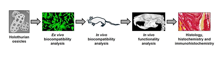
Abstract: Purpose: In the present study, we used holothurian ossicles (HOLO) extracted from sea cucumbers (holothurians) as novel biomaterials potentially useful in mandibular bone regeneration. Methods: HOLO particles were evaluated ex vivo and in vivo to determine biocompatibility and effectiveness in an animal model of bone defect. Results: First, ex vivo analyses found that HOLO were highly biocompatible when used with human cell cultures, as determined by LIVE/DEAD and DNA quantification assays, especially after 48 and 72 h of incubation. In contrast to control bone mineral particles (BP), cells cultured with HOLO tended to attach to these particles rather than to the culture surface, suggesting that the surface of HOLO could favor cell adhesion. In vivo analyses in Wistar rats showed that animals in which HOLO were grafted subcutaneously were devoid from any detectable side effects both at the systemic and local levels, and HOLO triggered a pro-regenerative M2-type macrophage response. When HOLO were applied in a model of mandibular bone defect, we found a positive effect of these particles as compared to negative controls, with a significant reduction of the size of the bone defect (3.36 ± 0.84 mm in HOLO vs. 9.16 ± 4.18 mm in controls) as determined by computed tomography (CT). Histologically, HOLO were associated to some ossification spots showing positive staining for toluidine blue, suggesting a process of osteoid formation, and an increased expression of osteonectin and osteocalcin, which were comparable or higher than control bone. Conclusions: These results suggest that HOLO could be safely used to induce mandible bone regeneration, and the use of these particles is associated to an increased bone regeneration process. Future studies should determine the clinical usefulness of these novel particles used in regenerative medicine. |
Keywords: Bone regeneration, holothurian particles, grafts, tissue engineering. |
Publication date: 14th February 2025 |
Copyright policy: © 2025 The Author(s). Published by Forum Multimedia Publishing, LLC. This article is distributed in accordance with Creative Commons Attribution Licence (http://creativecommons.org/licenses/by/4.0/). |
Article download: Pages 1-16 (PDF file) |